Final Project Update
Alterations in the extracellular matrix (ECM) structural components are a key feature of islet histopathology in type 1 diabetes (T1D). ECM is the non-cellular compartment of the tissue and is essential for the maintenance of tissue integrity and function. We recently found that hyaluronan (HA), a major component of the islet ECM, extensively accumulates in human islets in diabetes, forming large deposits along the islet microvessels and in the areas of islet inflammatory cell infiltrates defined as insulitis. The modification of the HA/ECM in islet tissue and in regions of islet inflammation matters because HA is a keystone molecule in the inflammatory milieu and has been increasingly implicated in the regulation of immune responses.
Click HERE to view the full project update.
Project Description
Type 1 diabetes (or T1D) is caused by a selective destruction of the body’s insulin-producing cells resulting from chronic inflammation of the pancreatic islet cells. The goal of this project is to discover, prior to patients exhibiting symptoms, the initial changes that take place in human islets, which are believed to cause insulin-producing cell loss at the onset of diabetes. These findings will then lead to an understanding of how this destructive process can be Blocked.
This project focuses on a specific tissue component named hyaluronan (or HA). Inflamed tissues have an abundance of HA which can influence the behavior of other cells. The intention is to determine how and why large amounts of HA are formed in human islets in the early stages of T1D. Early stages are characterized by the presence in blood of islet cell autoantibodies (or aAbs), a condition known as islet autoimmunity. Islet aAbs are used as markers to predict T1D.When T1D first starts, one type of islet cell aAb is measured in blood, but several years later, two or more types are detected. This project proposes that a modified HA-rich cell forms in human islets during the early stage of T1D when only one or two aAbs are detected. We believe that under the effect of inflammatory stimuli, islet cells make more HA, which then triggers insulitis and alters islet integrity and structure causing damage to the insulin-producing cells.
To answer the question of whether islets are actually modified and produce large amounts of HA in the early stage of T1D, we will examine a large set of pancreas tissues from non-diabetic organ donors who have tested positive for one or more islet cell aAbs. These pancreas tissues are identified through the Juvenile Diabetes Research Foundation network for Pancreatic Organ Donors.
Looking at the bigger picture, this research strives to develop a comprehensive understanding of the changes in islets which possibly cause islet inflammation and trigger insulitis in T1D. As such, this research will determine:
1) when the cell abnormalities first occur in the disease;
2) the mechanism that causes the abnormalities; and
3) how the abnormal cells result in T1D.
We will study human tissues; therefore, our new findings will be directly relevant to the human disease. This will affect patients with T1D, and help determine how to stop or prevent T1D.
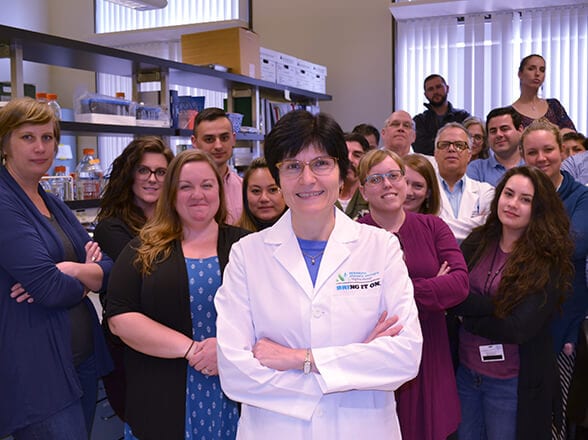
Working as a pathologist and diabetes researcher, Dr. Bogdani has built a broad and unique expertise in histopathology, high-resolution microscopy, quantitative pathology, islet biology and matrix biology, which is needed to carry out the proposed studies. This extensive expertise and the available institutional resources ensure project success.

