Where is He Now?
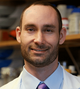
Todd Brusko, Ph.D. shares that his DRC-funded research project seeded key feasibility data that has been used for follow-on federal grants. He updated us on where he is at now. “DRC funding was critical support when I was starting as a young PI. I now head the research program within the UF Diabetes Institute.”
Final Project Report
In sum, the funding provided by the DRC was essential in jump starting this project and for generating the proof-of-concept data that will be needed for the next steps toward validation and clinical translation. We wish to extend our deepest gratitude to the donors and the support staff of the DRC for making this project a reality.
Project Description
Type 1 diabetes (T1D) results from the autoimmune destruction of pancreatic b cells. Prior animal model studies have demonstrated that a rare population of white blood cells, termed regulatory T cells (so called Tregs), can prevent and in certain contexts, even reverse the disease process immediately at onset. The field has addressed the challenges associated with how to isolate and grow these cells to therapeutic levels, yet there remain hurdles regarding how best to facilitate long-term engraftment of Tregs when returned to a patient.
The overall goal of our DRC supported project was to create a technology platform that would enable an optimized Treg cell therapy for the treatment of type 1 diabetes. Our technology involved the delivery of a key growth factor interleukin-2 (IL-2) to Treg. This growth factor was encapsulated in the form of nanoparticles (NP) to the surface of Tregs to facilitate extended release. Over the term of our award, we made significant progress toward the execution of our experimental aims.
The key milestones achieved, and outline of areas that require further development are summarized below:
- Manufacturing of IL-2 loaded NP – We hypothesized that the localized delivery of IL-2 to Tregs would be advantageous over the high doses of IL-2 that would need to be given systemically to restore immune regulation and support Treg cell engraftment. Therefore, we set out to manufacture biodegradable nanoparticles that would release the Treg growth and survival factor IL-2 for binding to Treg cell surfaces. For this purpose, we effectively manufactured NPs from the same material that FDA-approved dissolvable surgical sutures are manufactured from (known as PLGA). This approach resulted in nearly uniform creation of NPs (of ~300 nanometers) that could be effectively loaded with Treg cell growth factors. Our studies measured the efficiency of drug loading and release from these NPs.
- Conjugation of NP to Tregs – Once we had effectively manufactured NPs with our desired properties, we next set out to find a method to bind these particles to the surface of a living cell. Using a series of different surface chemistries, we were able to effectively bind NPs to the Treg cell surface and visualize this with microscopy. When NP were loaded with a fluorescent dye, we were able to quantify the number of NPs bound to the cell surface.
- Demonstration of biological activity of encapsulated IL-2—We next set out to determine if the IL-2 growth factor was still functional when conjugated to the surface of Tregs. To address these questions, we looked for markers of cell viability and signaling from the specific receptor molecule. These studies resulted in several essential findings; 1) the IL-2 loaded is active and effectively binds to the receptor of interest leading to downstream signaling, and 2) binding of IL-2 NPs was optimal for cell survival in comparison to NPs loaded with an irrelevant control protein.
- Animal models to demonstrate therapeutic efficacy—The ultimate test of our technology is whether it can positively impact a disease process. For our studies, we chose two different disease models in which to test our therapy. First, we set to test the effectiveness of mouse Tregs conjugated with IL-2 NPs in the context of the animal model of T1D, the non-obese diabetic (NOD) mouse. At the time of this writing, these studies are still ongoing. Secondly, we set out to test the effectiveness of human IL-2 conjugated NPs in an animal model of graft versus host disease. In this model, an immunodeficient mouse is engrafted human immune cells. Eventually, the human immune system recognizes the mouse tissues as “foreign” and begins to mount an inflammatory immune response against the host. In this system, Tregs play an important role in controlling this inflammation and tissue damage. Thus, we tested if IL-2 NPs conjugated to human Tregs would be more effective at controlling inflammation than NPs loaded with an irrelevant control protein. Excitingly, our initial data do support the notion of improved engraftment and function of IL-2 conjugated Tregs. These findings offer critical proof-of-principle data that our approach warrants further study and optimizations for therapeutic testing.
- Challenges and Future Directions— Our technology is designed to target and correct the underlying cause of autoimmune disease by restoring immune tolerance. Therefore, it is intended to impact the disease process in those at risk for T1D or who are close to onset and exhibit some residual b cell mass and function. Given our success loading bioactive IL-2, this project raises the potential to load and conjugate additional compounds or growth factors that might restore b cell mass and function. In this manner, Tregs with their inherent tissue homing capabilities could be used as living “smart drugs” to deliver critical compounds needed to impact the disease process in the pancreas.

