Final Project Update
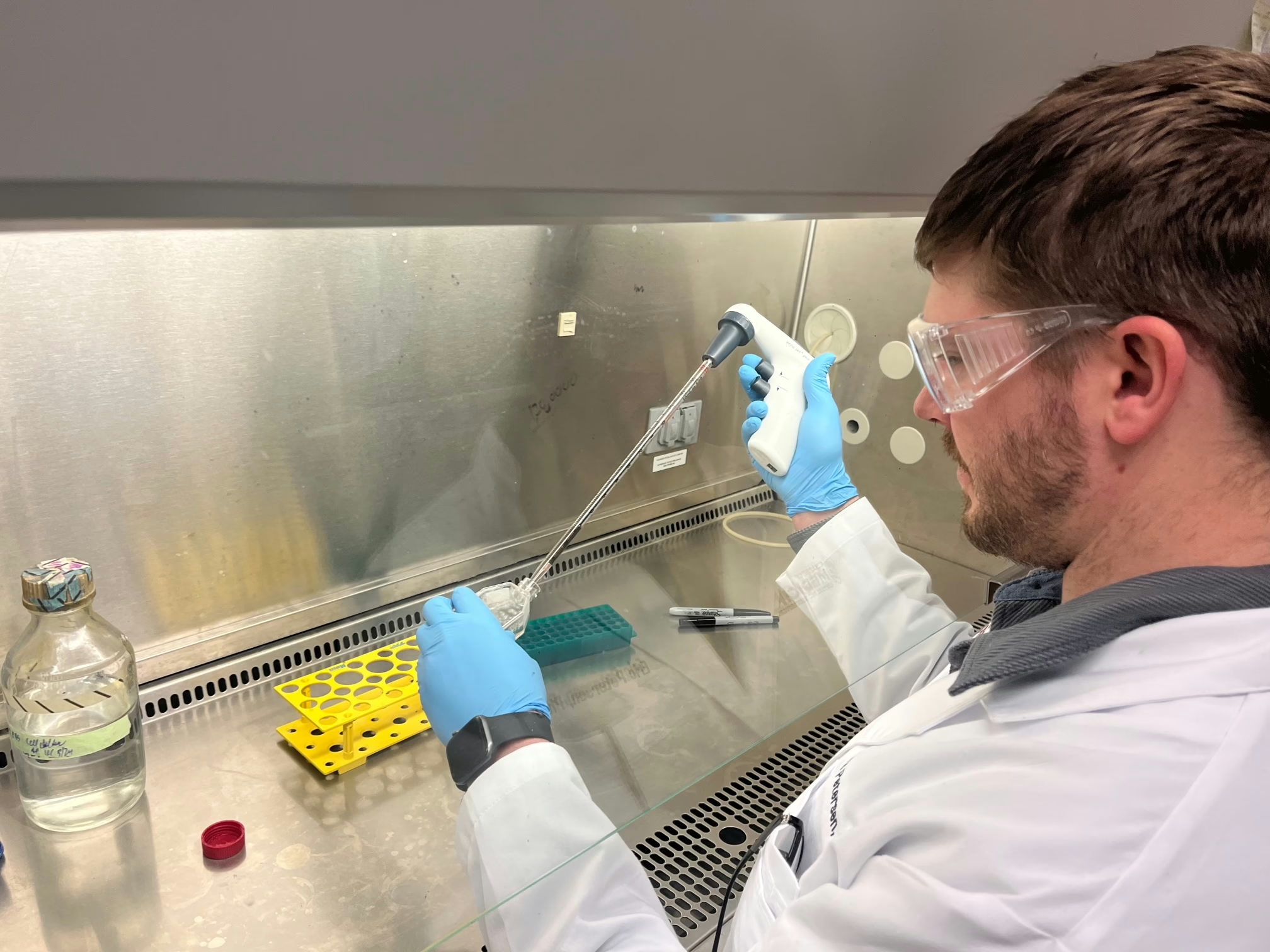
Thanks to DRC funding we managed to treat all 600 backcross mice necessary to ultimately map and identify genes uniquely contributing to immune checkpoint inhibition (ICI) induced type 1 diabetes elicited by anti-PD-1 treatment. A total of 301 anti-PD-1 treated mice and 301 PBS-treated controls have been entered into the study. To date, a total of 110 anti-PD-1 treated mice have gone diabetic compared to 31 PBS-controls (spontaneous diabetes). This corresponds to a diabetes rate of 36.5% in anti-PD-1 treated mice compared to 10.3% in PBS controls. The average age of onset for anti-PD-1 treated mice is at 14.3 weeks of age compared to 26.7 weeks of age for PBS-controls. Only 15 anti-PD-1 treated diabetic mice have gone diabetic after 17 weeks of age. As of the last month of funding, all remaining anti-PD-1 treated mice are beyond 17 weeks of age so additional diabetics will be few and far between. We will continue to monitor diabetes for all mice until they reach 40 weeks of age.
93 diabetic anti-PD-1 treated mice have been submitted for genotyping across 143,000 single nucleotide polymorphisms (SNPs) throughout the genome. 70 anti-PD-1 non-diabetic mice that have reached 40 weeks of age have also been submitted for genotyping. We anticipate in the coming days to obtain genotyping results for the final 24 of these mice required for an initial study to map genes uniquely contributing to ICI-induced diabetes. As soon as those results are obtained we will update the DRC with our findings. We have also submitted samples for 21 PBS-treated diabetic mice with additional mice to be submitted in the coming month. In order to map genes contributing to anti-PD-1 induced versus spontaneous diabetics, we will wait until the end of incidence in order to have the largest possible cohort sizes.
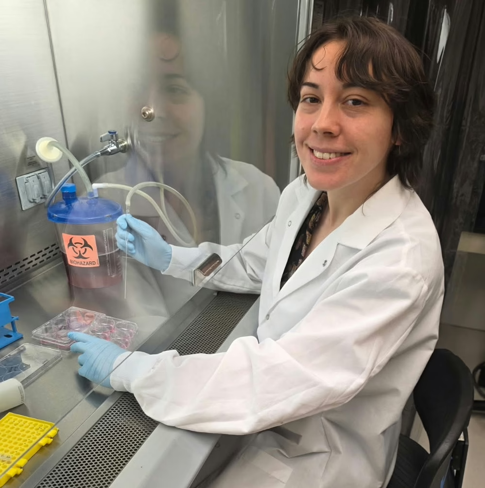
From an initial batch of 21 anti-PD-1 treated early diabetics (before 17 weeks of age), 7 anti-PD-1 treated late diabetics (after 17 weeks of age), 10 PBS-spontaneous diabetics, and 15 anti-PD-1 treated non-diabetics, RNA sequencing has now been performed on pancreatic lymph node CD8+ T-cells from a subset of 6-9 mice from each group. We are actively in the process of analyzing differential gene expression between the following comparisons: Anti-PD-1 Early diabetes vs Anti-PD-1 Late Diabetes; Anti-PD-1 Early diabetes vs Anti-PD-1 end of incidence Non-Diabetics; Anti-PD-1 Early diabetes vs PBS-spontaneous diabetes; and Anti-PD-1 Late Diabetes vs PBS-Spontaneous diabetes. We anticipate results of those comparisons in the coming week or two. We will update DRC with the results of those findings when they become available. We have continued to collect samples in the event mice have to be segregated into additional groupings for further analyses. As an example, we have banked an additional 31 samples of anti-PD-1 early diabetics since the initial RNA processing was performed.
Due to DRC funding, the data we have been able to obtain to date has allowed the larger Serreze laboratory at The Jackson Laboratory to secure follow-up funding on a larger project associated with ICI-induced autoimmunity. Once both initial and final mapping has been performed, our new funding will allow the creation of mouse models to further ICI-induced diabetes. Additionally, thanks to previous DRC funding (Gene-Specific Models and Therapies for Type One Diabetes) we had a murine-MHC-bare NOD mouse model that allows the introduction of human T1D-associated gene variants. After the completion of that project, we introduced the human T1D-associated HLA-DQ8 class II variant into our murine-MHC-bare mouse. Surprisingly, instead of the anticipated diabetes, mice developed myocarditis. Since myocarditis is another autoimmune side effect of ICI therapy, and this current DRC project provided proof-of-principal that there are genes associated specifically with anti-PD-1-induced autoimmunity, our new project will allow us to additionally assist patient populations that develop myocarditis after immune checkpoint therapy.
6-Month Project Update
Thanks to the Diabetes Research Connection, we have been able to ramp up the treatment of our mice with the immune checkpoint inhibitor (ICI) anti-PD-1 for mapping genes associated with ICI-induced diabetes. We have managed to double our treated mice in less than six months. Prior to DRC support, it took us a year and a half to accumulate an equivalent number of animals to our study. To date, 37/119 mice treated anti-PD-1 have gone diabetic, a rate of 31%. This rate has remained consistent throughout the study, confirming our initial hypothesis at the time of funding that we are looking at no more than two genes associated with ICI-induced diabetes. In contrast, our control group has had 12/159 mice go diabetic, a rate of ~7.5%. In addition to the enhanced frequency of diabetes occurring in our ICI-treated mice, there are also two phases of diabetes that have emerged. The average age of onset for ICI-induced diabetes is 15.9 weeks of age. In contrast, for spontaneous diabetes, the average age of onset is 22.8 weeks of age. Only 9 ICI-induced diabetic mice went diabetic over 16 weeks of age, and only 5 have gone diabetic over 20 weeks of age. Some of these mice likely represent “spontaneous” diabetics within the drug-treated group. To determine whether these mice are more similar to spontaneous or ICI-induced diabetics, we have decided to focus on RNA-sequencing (RNAseq) to allow gene expression profiles to “define” this phase of diabetes. Therefore, we have started to collect CD4 and CD8 T-cells from the pancreatic lymph nodes of diabetic spontaneous and ICI-induced diabetic mice. Mice that become diabetic over 20-weeks-of-age we are classifying as ICI-“late” to compare them to those ICI-induced diabetics that become diabetic prior to 20-weeks-of-age. We will be taking this same approach for mapping studies. As we obtain more mice within this window, we will be able to further refine the “cutoff” for group determination.
Due to the low frequency of diabetes in our spontaneous diabetes group, completing the mapping side of this project before the end of DRC funding was a longshot. We have now decided to define a fourth group of mice for RNAseq and mapping studies. Since 70% of ICI-treated mice remain diabetes-free, they represent an “internal control” group that initially we were not utilizing. Therefore, the first mapping will be between ICI-induced diabetics (diabetes before 20 weeks of age) to ICI-treated non-diabetics (40 weeks of age). To date, we have submitted and sequenced markers for 9 ICI-induced and 9 ICI-non-diabetics in addition to the parental strains that gave rise to these backcross mice. Within the next month, we will have submitted an additional 48 total samples for sequencing. Due to the large number of both ICI-induced and ICI-non-diabetics, we can obtain in this study, we now anticipate being able to have a preliminary mapping completed before the end of funding. As soon as we have managed to collect ~30 spontaneous diabetes, we can then perform the mapping comparison between the ICI-induced and spontaneous diabetics.
Project Description
Research Objective: Dr. Racine, an expert in immunology proposes to collaborate with fellow postdoc Dr. Jennifer Dwyer, an expert in molecular biology, to identify genes responsible for the fact some cancer patients undergoing immunotherapy treatment subsequently develop autoimmune T1D as a side effect. This research may lead to a method for blocking the onset of T1D, thereby preventing the disease.
A new sub-set of type 1 diabetes (T1D) patients have been identified within the field of cancer-immunotherapy. Immune checkpoint inhibitors (ICIs) are a relatively new class of therapeutics approved for treating a growing number of cancers including melanoma, non-small cell lung cancer, and Hodgkin’s lymphoma. While these therapies effectively treat the primary tumor, some patients can develop severe side effects, including the onset of T1D.
ICI-based therapies operate by activating white blood cells in order to attack tumors, but sometimes this process comes at the expense of also attacking normal tissue, including insulin-producing cells of the pancreas. To combat these deleterious side effects, patients are often prescribed drugs to suppress the immune response, making them susceptible to infection. With the increased use of ICI-based drugs, the number of patients developing autoimmune-type side effects is likely to rise.
Additionally, there is now evidence that a patient’s genes play a role in the success-rate of ICI therapy. It is believed that a patient’s genes could also play a role in whether they develop autoimmune side effects, like T1D. Researchers currently do not understand how close ICI-induced T1D resembles the “classical” form and onset of the disease. It is likely that many genes are shared between classical and ICI-induced T1D. Identification of the genes uniquely contributing to ICI-induced T1D should help improve the ability to predict patients who are likely to develop these side effects and thus develop better treatments. Understanding how these particular genes change the behavior of white blood cells may also provide important information for patients living with the classical form of T1D.
The Non-Obese Diabetic (NOD) mouse model develops T1D in a manner similar to human patients and thus provides a means to identify genes that contribute to T1D. We have made a novel modification to this model by replacing some mouse genes with the human counterparts that contribute to diabetes. These modifications, allow some of the mouse white blood cells that may cause T1D in humans to be identified. Using a standard genetic technique of selective breeding, we are currently mapping genes that uniquely contribute to ICI-induced T1D. Results to date indicate that there are likely no more than two genes uniquely contributing to ICI-induced T1D.
This project aims to expand upon our preliminary studies to obtain appropriate numbers of ICI-treated and control NOD mice (~300 each) in order to map the genes distinguishing ICI-induced and spontaneous T1D. We will examine differences in the white blood cells between the two types of T1D.
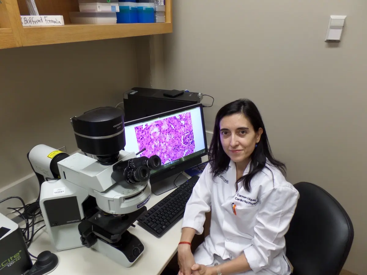
The Serreze laboratory has a long history of identifying genes contributing to T1D development. Once the gene-regions in question have been identified, with the help of experts in the Jackson Laboratory, we propose to test candidate genes within these regions using CRISPR/Cas9 technology. This will aid in determining how manipulation of such genetic pathways affects classical spontaneous and ICI-induced T1D development.