DRC & Research News

This page shares the latest news in T1D research and DRC’s community.

Get the most recent diabetes research news, delivered straight to your inbox

Antibodies in the blood effect Diabetes

Early Signs in Young Children Predict Type 1 Diabetes

New research shows that it is possible to predict the development of type 1 diabetes. By measuring the presence of autoantibodies in the blood, it is possible to detect whether the immune system has begun to break down the body’s own insulin cells.

“In the TEDDY study we have found that autoantibodies often appear during the first few years of life,” said Professor Åke Lernmark from Lund University, who is leading the study in Sweden.

The TEDDY study, funded by the US National Institutes of Health (NIH), involves 8 600 children from Sweden, the US, Germany and Finland. The children have an increased hereditary risk of type 1 diabetes, detected at birth through tests on blood from the umbilical cord. TEDDY stands for “The Environmental Determinants of Diabetes in the Young.”

Antibodies are part of the body’s immune system and the presence of antibodies in the blood is a sign that the immune system has reacted to an intruder such as a virus or a bacteria. Sometimes, the immune system mutinies and attacks the body. Autoantibodies are a sign of an autoimmune disease and form markers indicating that an attack is underway, for example on the body’s own insulin cells.

The new findings from the TEDDY study have been published in the journal Diabetologia and show that there are three ways to predict the development of type 1 diabetes.

Three ways to predict development of type 1 diabetes:

1. If the autoantibody first discovered attacks insulin (IAA) In Sweden this usually takes place at the age of 18 months. However, in the study as a whole most babies affected were less than a year old.

“If a second autoantibody is detected later, then the person will get diabetes — but it may take up to 20 years,” said Åke Lernmark.

2. If the first autoantibody targets GAD65 (GADA), a protein inside the insulin-producing cells In Sweden this usually happens at the age of two and half, whereas in the study as a whole it was most common at the age of two.

3. If both autoantibodies are first found together

“In TEDDY, 40 per cent of these children had already developed diabetes,” said Åke Lernmark

Of the participating children, 6.5 per cent had their first autoantibody before the age of six.

  • In 44 per cent of cases, they only had an autoantibody against insulin (IAA). Most of them had this by the age of 1-2.
  • In 38 per cent of cases, GAD65 autoantibodies (GADA) were detected. The numbers increased until the age of two and then remained constant.
  • In 14 per cent of cases both autoantibodies were found at the same time, with a peak at the age of 2-3.

The hereditary risk of type 1 diabetes determined which autoantibody the children had. However, it is still not known what causes the immune system to start attacking the body’s own insulin cells to start with. One theory is that a viral infection could be the trigger.

“It is possible that there are two different diseases involved. Perhaps one virus triggers the autoantibodies against insulin and another one the autoantibodies against GAD65,” said Åke Lernmark.

Footnote: Since the birth of the children in the TEDDY study, their parents have kept regular, detailed food diaries, submitted blood and stool samples, nail samples and information about illnesses and medication. When autoantibodies are detected in a child’s blood, the researchers begin the sizeable task of analysing all the material in the hunt for what it is that may have caused the immune system to mutiny.


Lund University. “Early signs in young children predict type 1 diabetes.” ScienceDaily. ScienceDaily, 26 February 2015. Click here to read the originally published article on Science Daily.


Learn More +
Protein Sheild

Protein That Repels Immune Cells Protects Transplanted Pancreatic Islets From Rejection

Protein SheildTransplanting islets encapsulated with CXCL12 restores blood sugar control without immunosuppression in animal models of diabetes

An approach developed by Massachusetts General Hospital (MGH) investigators may provide a solution to the limitations that have kept pancreatic islet transplantation from meeting its promise as a cure for type 1 diabetes. In the March issue of the American Journal of Transplantation, the research team reports that encapsulating insulin-producing islets in gel capsules infused with a protein that repels key immune cells protected islets from attack by the recipient’s immune system without the need for immunosuppressive drugs, restoring long-term blood sugar control in mouse models. The technique was effective both for islets from unrelated mice and for islets harvested from pigs.

“Protecting donor islets from the recipient’s immune system is the next big hurdle toward making islet transplantation a true cure for type 1 diabetes,” says Mark Poznansky, MD, PhD, director of the MGH Vaccine and Immunotherapy Center, who led the study. “The first was generating enough insulin-producing islets, which has been addressed by several groups using pig islets or – as announced last fall by Doug Melton’s team at the Harvard Stem Cell Institute – with islet cells derived from human stem cells. Now our technology provides a way to protect islets or other stem-cell-derived insulin-producing cells from being destroyed as soon as they are implanted into a diabetic individual without the need for high-intensity immunosuppression, which has its own serious side effects.”

While transplantation of pancreatic islets has been investigated for several decades as a treatment and potential cure for type 1 diabetes, its success has been limited. Along with the risk of rejection that accompanies all organ transplants – a risk that is even greater for cross-species transplants – donated islets are subject to the same autoimmune damage that produced diabetes in the first place. The immunosuppressive drugs used to prevent organ rejection significantly increase the risk of infections and some cancers, and they also can contribute directly to damaging the islets. Among the strategies investigated to protect transplanted islets are enclosing them in gel capsules and manipulating the immune environment around the implant. The MGH-developed approach includes aspects of both approaches.

Previous research from the MGH team demonstrated that elevated expression of a chemokine – a protein that induces the movement of other cells – called CXCL12 repels the effector T cells responsible for the rejection of foreign tissue while attracting and retaining regulatory T cells that suppress the immune response. For the current study they investigated how either coating islets with CXCL12 or enclosing them in CXCL12 gel capsules would protect islets transplanted into several different mouse models.

Their experiments revealed that islets from nondiabetic mice, either coated with CXCL12 or encapsulated in a CXCL12-containing gel, survived and restored long-term blood sugar control after transplantation into mice with diabetes that was either genetically determined or experimentally induced. CXCL12-encapsulated islets were even protected against rejection by recipient animals previously exposed to tissue genetically identical to that of the donor, which usually would sensitize the immune system against donor tissue. CXCL12-encapsulated pig islets successfully restored blood sugar control in diabetic mice without being rejected. The ability of CXCL12 – either as a coating or encapsulating gel – to repel effector T cells and attract regulatory T cells was also confirmed.

“While studying this procedure in larger animals is an essential next step, which is currently underway with the support of the Juvenile Diabetes Research Foundation, we expect that this relatively simple procedure could be readily translatable into clinical practice when combined with technologies such as stem-cell-derived islets or other insulin-producing cells and advanced encapsulation devices,” says Poznansky, an associate professor of Medicine at Harvard Medical School. “We also hope that CXCL12 will have a role in protecting other transplanted organs, tissues and cells as well as implantable devices, a possibility we are actively investigating.”

http://www.medicalnewstoday.com/releases/289590.php?tw


Learn More +
MRI

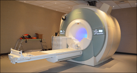
Scientists Use MRI to Visualize Pancreas Inflammation in Type 1 Diabetes

MRIA pilot study led by researchers at Joslin Diabetes Center has revealed that it is possible to use magnetic resonance imaging (MRI) to “see” the inflammation in the pancreas that leads to type 1 diabetes. This discovery could be a boon for research on methods to slow or halt the disease at an early stage, and could also guide insights into how diabetes progresses.

This clinical study, published in the Proceedings of the National Academy of Sciences, tested the possibility of imaging inflammation in the pancreas of human volunteers using ferumoxytol, a coated iron nanoparticle approved by the FDA as an iron replacement therapy, and MRI. Ferumoxytol leaks out of blood vessels in areas of inflammation and is taken up by immune cells called macrophages, which congregate at sites of inflammation. (AMAG Pharmaceuticals, which markets ferumoxytol under the name Feraheme®, was not involved in the study .)

Autoimmunity and inflammation directed against the pancreas and its insulin-producing beta cells underlie the development of type 1 diabetes. However, while tests for autoantibodies (antibodies against the pancreas) can reveal whether a patient’s immune system has at some point attacked the pancreas, these antibodies are not always a good marker for predicting whether a given individual will develop full-blown diabetes.

“Many people have genetic variants that put them at risk for type 1 diabetes,” explains study co-lead author Jason Gaglia, M.D., M.M.Sc., an Assistant Investigator in the Section of Immunobiololgy at Joslin. “Some develop autoimmunity, but only a small number develop clinical disease.”

In addition, the development of therapies that could potentially halt patients’ progression from pancreatic inflammation to diabetes has been hampered by the long lead times needed in order to tell whether a given therapy has an effect.

“If you want to study in people an immunodulatory agent right now, for diabetes it takes years,” Gaglia says. He explains that the end measurements for whether such therapies work relate directly to pancreatic function, changes in which may not become apparent for a long time. With imaging, he continues, “you could have an answer in a matter of months.”

For the study, Gaglia and his collaborators, including study co-lead author Mukesh Harisinghani, M.D., of Massachusetts General Hospital, recruited 11 patients with newly diagnosed type 1 diabetes and evidence of antibodies against the pancreas, a sign that their beta cells were under inflammatory attack. They also recruited 10 controls with no sign or family history of diabetes.

To visualize inflammation across the whole pancreas, the researchers adapted MRI mapping algorithms originally developed for whole brain scanning. All other components of the experiment, including MRI equipment and ferumoxytol, are widely available. Gaglia notes that the ferumoxytol dose used in this imaging study is approximately one quarter of the dose used therapeutically for iron replacement. “These are all off-the-shelf components,” he says. “Other centers can do this now.”

Ferumoxytol-MRI images of the patient group showed clear evidence of ferumoxytol accumulation in the pancreas, indicating ongoing inflammation. By comparison, images from the control group did not.

The researchers believe this imaging technique could have a range of applications in diabetes research and help build a better understanding of the natural history of type 1 diabetes. Already, Gaglia says, the ferumoxytol-MRI images reveled that in their patient group, “inflammation was not uniform across the entire pancreas. There was also a large amount of variation between individuals, which aligns with what you see clinically. That’s never been shown in living humans before.”

This imaging approach could also, in the future, help better define which patients with autoimmunity will likely progress to diabetes and classify subgroups of patients who might benefit from different therapeutic strategies. It could also identify those patients with early signs of autoimmunity who might be good candidates for clinical research studies.

“Only about five percent of the first degree family members of a person with diabetes will develop occult disease,” Gaglia, who is firm that for the moment this imaging technique should only be used in the context of research. “It might make sense to scan people in that group to see who is likely to progress and who isn’t. Those who are progressing may be the ones you would want to recruit for research on immunomodulatory therapies.”

Other contributors to this PNAS study include Iman Aganj, Gregroy Wojtkiewicz, Sandeep Hedgire, and Ralph Weissleder of Massachusetts General Hospital; and Christophe Benoist and Diane Mathis of Harvard Medical School.


Story Source:

The above story is based on materials provided by Joslin Diabetes CenterNote: Materials may be edited for content and length.


Journal Reference:

  1. Jason L. Gaglia, Mukesh Harisinghani, Iman Aganj, Gregory R. Wojtkiewicz, Sandeep Hedgire, Christophe Benoist, Diane Mathis, Ralph Weissleder.Noninvasive mapping of pancreatic inflammation in recent-onset type-1 diabetes patientsProceedings of the National Academy of Sciences, 2015; 201424993 DOI: 10.1073/pnas.1424993112

Cite This Page:

Joslin Diabetes Center. “Scientists use MRI to visualize pancreas inflammation in type 1 diabetes.” ScienceDaily. ScienceDaily, 18 February 2015. <www.sciencedaily.com/releases/2015/02/150218123907.htm>.


Learn More +
Lucentis Bottle

FDA Approves Lucentis for Treatment of Diabetic Retinopathy in People With Diabetic Macular Edema

Lucentis BottleGenentech, a member of the Roche Group, has announced that the U.S. Food and Drug Administration (FDA) approved Lucentis (ranibizumab injection) for the treatment of diabetic retinopathy (DR) in people with diabetic macular edema (DME). DME impacts nearly 750,000 Americans, about 10 percent of people with DR.

The FDA granted Lucentis Breakthrough Therapy Designation and Priority Review for this indication based on results from the RISE and RIDE Phase III clinical trials.

“While there are various options for treating diabetic macular edema, before today none were approved showing improvement in retinopathy,” said Sandra Horning, M.D., chief medical officer and head of Global Product Development. “With today’s approval, people with diabetic macular edema now have a FDA-approved medicine that showed meaningful improvements in retinal damage from diabetes, in addition to the established improvement in vision.”

Almost 29 million Americans have diabetes. The longer people have diabetes, especially if it is poorly managed, the higher their risk for developing DR. It is caused by elevated blood sugar levels damaging the fine blood vessels of the retina, the light-sensitive tissue at the back of the eye necessary for good vision.

DR with DME is a common diabetic eye disease and a leading cause of blindness in American adults under 55.1 DR with DME can lead to conditions that threaten vision.

The FDA designates Breakthrough Therapy to a medicine if it is intended to treat a serious or life-threatening disease and if preliminary clinical research suggests it may provide substantial improvement on clinically significant endpoints over existing therapies.

The FDA grants Priority Review to medicines that, if approved, would have the potential to provide significant improvements in the safety or effectiveness of the treatment, diagnosis, or prevention of serious conditions when compared to standard applications.

In 2012, Lucentis was the first medicine approved by the FDA for the treatment of DME. Lucentis has also been an important option for patients with wet age-related macular degeneration (wet AMD) since 2006 and macular edema following retinal vein occlusion (RVO) since 2010.

About RISE and RIDE

RISE and RIDE are two identically-designed, parallel, double-masked, sham treatment-controlled trials in 759 patients with DR and DME at baseline who were randomized into three groups to receive monthly treatment with 0.3 mg Lucentis, 0.5 mg Lucentis or sham injection. The primary outcome in RISE and RIDE was visual acuity gain at 24 months for DME patients.

The safety and efficacy of Lucentis for the treatment of DR with DME was assessed over three years in patients with baseline DR severity scores ranging from 10 to 75 in the study eye (on the ETDRS diabetic retinopathy severity scale). Secondary and exploratory outcomes were evaluated at 24 months. At Month 24, a higher proportion of patients had observed a three-step or better improvement of their disease compared to sham, as determined by color fundus photography. The safety in the RISE and RIDE Phase III trials was consistent with previous studies.

In the third year of the studies, patients from the control group had the option to cross over to receive monthly treatment with 0.5 mg Lucentis; patients originally randomized to 0.3 mg or 0.5 mg Lucentis continued to receive the same dose and all patients were followed for 12 additional months. The 0.3 mg dose of Lucentis is approved for both DME and for DR in people with DME.

About Lucentis

Lucentis is a vascular endothelial growth factor (VEGF) inhibitor designed to bind to and inhibit VEGF-A, a protein that is believed to play a critical role in the formation of new blood vessels (angiogenesis) and the hyperpermeability (leakiness) of the vessels.

Lucentis is FDA-approved for the treatment of wet AMD, macular edema following RVO, DME and DR in people with DME. Genentech has conducted eight key clinical trials with Lucentis. The medicine has been studied in 21 clinical trials worldwide in more than 9,080 patients.

Lucentis was developed by Genentech. The company retains commercial rights in the U.S. and Novartis has exclusive commercial rights for the rest of the world.

Outside the U.S., Lucentis is approved in more than 100 countries to treat patients with wet AMD, for the treatment of DME, and due to macular edema secondary to both branch retinal vein occlusion (BRVO) and central retinal vein occlusion (CRVO).

Lucentis Important Safety Information

Patients should not use Lucentis if they have an infection in or around the eye or are allergic to Lucentis or any of its ingredients. Lucentis is a prescription medication given by injection into the eye and it has side effects. Some Lucentis patients have had detached retinas and serious infections inside the eye.

Uncommonly, Lucentis patients have had serious, sometimes fatal problems related to blood clots, such as heart attacks or strokes.

Some patients have had increased eye pressure before and within one hour of an injection.

Serious side effects include inflammation inside the eye and, rarely, problems related to the injection procedure such as cataracts. These side effects can make vision worse.

The most common eye-related side effects are increased redness in the white of the eye, eye pain, small specks in vision and increased eye pressure. The most common non-eye-related side effects are nose and throat infections,headache, lung/airway infections, and nausea.

If the eye becomes red, sensitive to light, or painful, or if there is a change in vision, patients should call or visit an eye doctor right away.

Lucentis is for prescription use only.

For additional safety information, please see Lucentis full prescribing information, available here:http://www.gene.com/download/pdf/lucentis_prescribing.pdf

Adapted by MNT from original media release

http://www.medicalnewstoday.com/releases/289246.php?tw


Learn More +
Blood Prick

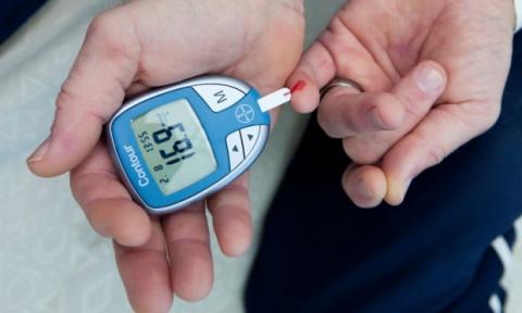
‘Smart’ Insulin May Ease Burden of Type 1 Diabetes Patients, Research Suggests

Blood PrickDiabetes patients could be spared the burden of constantly monitoring blood sugar levels after scientists have developed a “smart” form of insulin.

The new compound, which was shown to be effective in mice, automatically activates when blood sugar levels soar, and remains in circulation for up to 24 hours. In the future, patients could inject the insulin once a day, or even less frequently, overcoming the need for constant self-monitoring and insulin top-ups after meals.

Danny Chou, a chemical biologist who led the research at the University of Utah, said: “Diabetic patients still need to guess to some extent how much insulin they need. With this you would just inject it and it wouldn’t matter if you overshot because its activity would stop when glucose levels get too low.”

About 400,000 people in Britain have type 1 diabetes, including 30,000 children. The condition, which normally begins in childhood, is an autoimmune disease in which the body kills off all its pancreatic beta cells. The cells produce insulin, which regulates blood sugar, and without beta cells, the body’s sugar levels fluctuate wildly, meaning that patients need to monitor glucose and typically inject insulin several times each day.

Taking too much insulin can drive blood sugar levels too low, leading to hypoglycaemia– such episodes are responsible for around 10% of deaths in type 1 diabetes.

Taking too little insulin means blood sugar levels are consistently too high, which can lead to serious complications in the long term, such as blindness and nerve damage. “In theory, with this there would be none of these glucose problems,” said Dr Chou.

The smart insulin, known as Ins-PBA-F, is engineered to bind to a blood protein, called albumin, as soon as it is injected. This means it is essentially stored in a reservoir. The insulin is then released when blood sugar levels reach a certain threshold, meaning it can act to lower the circulating glucose.

In the study, published on Monday in the Proceedings of the National Academies of Science, diabetic mice given a single daily injection of the modified insulin had glucose levels that were as stable as those seen in healthy mice. The team hope to test the treatment in patients within two to five years.

Karen Addington, chief executive of the Juvenile Diabetes Research Foundation in the UK, said: “For many people living with type 1 diabetes, achieving good blood glucose control is a daily battle. A smart insulin would eliminate hypos – which are what many with type 1 diabetes hate most. It would enable people with type 1 diabetes to achieve near perfect glucose control, all from a single injection per day or even per week. That’s really exciting.”

Dr Richard Elliott, of Diabetes UK, said the approach had the potential to make it easier for people with diabetes to manage their condition, but cautioned that the progress towards clinical versions of the treatment could be slow. “Years of further research and clinical trials will be needed to find out if a similar drug could be used safely and effectively by people with diabetes,” he said.


http://www.theguardian.com/society/2015/feb/09/smart-insulin-may-ease-bu…


Learn More +
Pediatrician with Diabetic Child

Change in Gut Bacteria May Precede Type 1 Diabetes in Kids

Pediatrician with Diabetic ChildTHURSDAY, Feb. 5, 2015 (HealthDay News) — In some young children who develop type 1 diabetes, a change in normal stomach bacteria can precede the disease by a year, a small study has found.

The findings, published Feb. 5 in the journal Cell, Host & Microbe, are based on just 33 children at increased genetic risk of type 1 diabetes. And experts stressed that it’s too early to tell what it all could mean.

But one hope is that the results will lead to an early diagnostic test for type 1 diabetes, said researcher Aleksandar Kostic, a postdoctoral fellow at the Broad Institute of MIT and Harvard.

There is also the possibility of developing new therapies for type 1 that would target the “ecosystem” of the gut, he said.

But that would be a long way off, Kostic stressed. “These findings open up a promising new avenue for more research,” he said. “But that’s all we can say for now.”

Type 1 diabetes differs from the much more common type 2 diabetes, which is often linked to older age and obesity. In type 1, the immune system mistakenly kills off pancreatic cells that make the blood-sugar-regulating hormone insulin. To survive, people with type 1 have to take frequent injections of insulin, or use an insulin pump, for the rest of their lives.

As many as 3 million Americans have type 1 diabetes, according to the JDRF (formerly Juvenile Diabetes Research Foundation), a New York-based organization that funds research into the disease. Often, the disease arises in childhood, but there are adult-onset cases, too.

Scientists do not know exactly what causes the abnormal immune reaction. But people who carry certain gene variants related to immune system function have a higher-than-normal risk of developing type 1 diabetes.

The new study followed 33 babies from Finland and Estonia who carried some of those gene variants. Kostic and his colleagues analyzed stool samples from the children to chart changes in the trillions of bacteria, viruses and other microorganisms that dwell in the gut — what scientists call the “microbiome.”

By age 3 years, four children had developed type 1 diabetes. And those children showed a clear change in gut “bugs” about one year before the onset of the disease.

“What we saw were huge alterations,” Kostic said. “That included a drop in the overall diversity of the ‘community.'”

He likened the situation to a rain forest that is being cleared. The decline in its natural diversity opens the door for some “bad players” to take root.

In this case, children who went on to develop diabetes showed a decline in “good bugs” that produce beneficial fatty acids, and an increase in organisms linked to inflammation, Kostic explained.

But it’s not clear, he said, whether that shift in the gut helps cause the abnormal immune reaction behind type 1 diabetes or results from it. That’s a question for future research, Kostic said.

Another expert agreed. A big next step will be to understand the physiological “pathways” that the gut microorganisms affect, said Jessica Dunne, director of discovery research for JDRF, which funded the study.

“We’re still a long way off from a therapy,” Dunne said. But, she added, researchers are already interested in whether a “probiotic” therapy might help prevent or delay type 1 diabetes in children at increased risk. Probiotics are live bacteria like those found naturally in the human body.

Another question, Kostic said, is whether these findings in toddlers would be true of older children or adults who develop type 1 diabetes. He said it’s fairly uncommon for the disease to be diagnosed by age 3, and it’s possible there’s something “unique” about children who develop it that early.

Dunne agreed that the disease process could be different in other age groups.

If a change in gut bacteria is a sign of diabetes to come, that could offer a way to catch the disease process early, Kostic and Dunne said.

However, only a small minority of kids who carry susceptibility genes actually develop type 1 diabetes, Kostic noted. “So genetics doesn’t tell you much,” he said.

Dunne said researchers would like to develop some way to predict which kids seem to be “on the path” to type 1 diabetes. That will be needed if any therapies to prevent or delay the disease become available.


SOURCES: Aleksandar Kostic, Ph.D., postdoctoral fellow, Broad Institute of MIT and Harvard, Cambridge, Mass.; Jessica Dunne, Ph.D., director, discovery research, JDRF, New York City; Feb. 5, 2015, online, Cell, Host & Microbe

http://consumer.healthday.com/diabetes-information-10/misc-diabetes-news…


Learn More +
Fruit Fly Used in Research

Stanford Researchers Discover Insulin-Decreasing Hormone in Flies, Humans

Fruit Fly Used in ResearchAn insulin-regulating hormone that, until now, only had been postulated to exist has been identified by researchers at the Stanford University School of Medicine.

The hormone, called limostatin after the Greek goddess of starvation, Limos, tamps down circulating insulin levels during recovery from fasting or starvation. In this way, it ensures that precious nutrients remain in the blood long enough to rebuild starving tissues, rather than being rapidly squirreled away into less-accessible fat cells.

The researchers first discovered limostatin in fruit flies but then quickly identified a protein with a similar function in humans.

“Starvation or famine is an ancient, ever-present specter faced by all living organisms,” said Seung Kim, MD, PhD, professor of developmental biology. “The ways to deal with it metabolically are likely to be ancient and conserved. This research clearly connects the dots between flies and humans, and identifies a new potential way to regulate insulin output in humans.”

In particular, members of a family with an inherited mutation in the human analog of limostatin exhibited many of the same physiological characteristics as flies genetically engineered to be unable to produce limostatin — namely, high levels of circulating insulin, low blood sugar levels and a tendency toward early onset obesity.

A paper describing the research findings was published in Cell Metabolism. Kim is the senior author, and graduate student Ronald Alfa is the lead author.

The metabolic dance

Insulin is a key player in the complicated metabolic dance shared by nearly all organisms. Its importance can hardly be overstated. After a meal, animals and humans produce insulin in response to the increase in blood sugars that occur as a meal is digested. This insulin stimulates the storage of circulating sugars into muscle and fat cells for future use. Too little insulin, or an inability of the body to respond correctly to its signal, causes a dangerous spike in blood sugar levels. Conversely, too much insulin can cause a rapid drop. Both conditions can be life-threatening, and ongoing swings in blood sugar levels can lead to complications, such as blindness, poor circulation and kidney failure.

“This work has critical ramifications for our understanding of metabolism, and has the potential to transform our approach to treating diseases like diabetes,” said Domenico Accili, MD, director of the Columbia University Diabetes and Endocrinology Research Center and Columbia’s Russell Barry Foundation Professor of Diabetes.

“The discovery of limostatin, a new hormone that can act to decrease insulin release, is an important advance,” added Accili, who was not involved with the research. “The notion that mammals express a related family of intestinal hormones that can affect insulin secretion may inform new efforts to find drugs that combat diabetes in humans.”


http://www.medicalnewstoday.com/releases/288946.php?tw


Learn More +
unmethylated DNA Affects Diabetes

A Simple Method to Monitor β Cell Death in Individuals At-Risk for Type 1 Diabetes

unmethylated DNA Affects DiabetesType 1 diabetes is characterized by death and reduced function of β cells, which produce insulin.

The presence of specific autoantibodies can identify individuals at risk of developing type 1 diabetes, and many of these at-risk individuals exhibit evidence of β cell death before the onset of clinical symptoms. However, the course of β cell death that ultimately leads to the development of type 1 diabetes is poorly understood.

A new study in the Journal of Clinical Investigation reveals that a DNA biomarker can be used to evaluate the extent of β cell death and type 1 diabetes risk. Kevan Herold and colleagues at Yale University measured the amount of unmethylated insulin DNA in blood in healthy individuals and a cohort at risk of developing type 1 diabetes.

Insulin DNA is methylated in most cell types, which prevents its expression. Therefore, the authors predicted that the unmethylated form of DNA would only be present if there had been substantial β cell death and lysis.

At-risk individuals had much higher levels of unmethylated insulin DNA, and the increase in this DNA biomarker also correlated with a reduction in other measures of β cell function. Moreover, the levels of unmethylated DNA further increased in the period prior to clinical onset.

Together, these results of this study indicate that unmethylated insulin DNA can be used as a marker of β cell death.


http://www.medicalnewstoday.com/releases/288897.php?tw

Title: β Cell death and dysfunction during type 1 diabetes development in at-risk individuals


Learn More +
Insulin inhaler

Inhaled Insulin Powder Now Available in Pharmacies

PInsulin inhalerARIS and VALENCIA, Calif. — Afrezza, an inhaled insulin powder, is now available by prescription in U.S. retail pharmacies, Sanofi and MannKind Corp. announced. The drug is approved by the Food and Drug Administration to control high blood sugar in adults who have type 1 and type 2 diabetes.
Afrezza consists of a dry formulation of human insulin and is administered via a small, portable inhaler to help patients control blood sugar levels. The drug — which the companies said is rapidly absorbed, with a short duration of action — is administered before patients consume a meal.
“Many people living with diabetes are not able to control their blood sugar on their current medications and may benefit from using insulin. Now they have another option to administer insulin that is not an injection,” said Dr. Janet McGill, M.D., professor of medicine at Washington University School of Medicine in St. Louis and Afrezza clinical trial investigator. “This delivery option may help change the dialogue between health care professionals and people living with diabetes about initiating or intensifying insulin therapy.”
The companies noted that Afrezza cannot be used as a treatment for diabetic ketoacidosis, nor should it be used in patients suffering from chronic lung diseases.
Learn More +

OUR PROJECTS

See our approved research projects and campaigns.

Role of the integrated stress response in type 1 diabetes pathogenesis
In individuals with type 1 diabetes (T1D), the insulin-producing beta cells are spontaneously destroyed by their own immune system. The trigger that provokes the immune system to destroy the beta cells is unknown. However, accumulating evidence suggest that signals are perhaps first sent out by the stressed beta cells that eventually attracts the immune cells. Stressed cells adapt different stress mitigation systems as an adaptive response. However, when these adaptive responses go awry, it results in cell death. One of the stress response mechanisms, namely the integrated stress response (ISR) is activated under a variety of stressful stimuli to promote cell survival. However, when ISR is chronically activated, it can be damaging to the cells and can lead to cell death. The role of the ISR in the context of T1D is unknown. Therefore, in this DRC funded study, we propose to study the ISR in the beta cells to determine its role in propagating T1D.
Wearable Skin Fluorescence Imaging Patch for the Detection of Blood Glucose Level on an Engineered Skin Platform
zhang
A Potential Second Cure for T1D by Re-Educating the Patient’s Immune System
L Ferreira
Validating the Hypothesis to Cure T1D by Eliminating the Rejection of Cells From Another Person by Farming Beta Cells From a Patient’s Own Stem Cells
Han Zhu
Taming a Particularly Lethal Category of Cells May Reduce/Eliminate the Onset of T1D
JRDwyer 2022 Lab 1
Can the Inhibition of One Specific Body Gene Prevent Type 1 Diabetes?
Melanie
Is Cholesterol Exacerbating T1D by Reducing the Functionality and Regeneration Ability of Residual Beta Cells?
Regeneration Ability of Residual Beta Cells
A Call to Question… Is T1D Caused by Dysfunctionality of Two Pancreatic Cells (β and α)?
Xin Tong
Novel therapy initiative with potential path to preventing T1D by targeting TWO components of T1D development (autoimmune response and beta-cell survival)
flavia pecanha