DRC & Research News

This page shares the latest news in T1D research and DRC’s community.

Get the most recent diabetes research news, delivered straight to your inbox

MIT-Diabetes-Stem-1_0

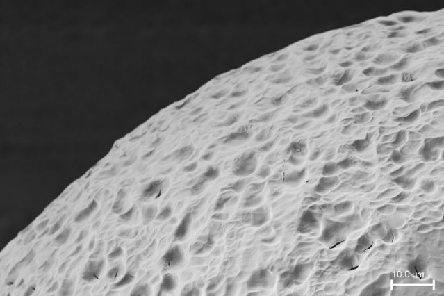
No More Insulin Injections?

Original article written by Anne Trafton for MIT News Office on January 26, 2016. Click here to read the original article.

MIT-Diabetes-Stem-1_0
A stealth material surface, shown here, has been engineered to provide an “invisibility cloak” against the body’s immune system cells. In this electron microscopy image, you can see the material’s surface topography.
Courtesy of the researchers.

In patients suffering from Type 1 diabetes, the immune system attacks the pancreas, eventually leaving patients without the ability to naturally control blood sugar. These patients must carefully monitor the amount of sugar in their blood, measuring it several times a day and then injecting themselves with insulin to keep their blood sugar levels within a healthy range. However, precise control of blood sugar is difficult to achieve, and patients face a range of long-term medical problems as a result.

A better diabetes treatment, many researchers believe, would be to replace patients’ destroyed pancreatic islet cells with healthy cells that could take over glucose monitoring and insulin release. This approach has been used in hundreds of patients, but it has one major drawback — the patients’ immune systems attack the transplanted cells, requiring patients to take immunosuppressant drugs for the rest of their lives.

MIT-Diabetes-Stem-2
Glucose-stimulated insulin-producing cells, derived from stem cells, are protected inside capsules that are engineered to be invisible to the host immune system.
Courtesy of the researchers.

Now, a new advance from MIT, Boston Children’s Hospital, and several other institutions may offer a way to fulfill the promise of islet cell transplantation. The researchers have designed a material that can be used to encapsulate human islet cells before transplanting them. In tests on mice, they showed that these encapsulated human cells could cure diabetes for up to six months, without provoking an immune response.

Although more studies are needed, this approach “has the potential to provide diabetics with a new pancreas that is protected from the immune system, which would allow them to control their blood sugar without taking drugs. That’s the dream,” says Daniel Anderson, the Samuel A. Goldblith Associate Professor in MIT’s Department of Chemical Engineering, a member of MIT’s Koch Institute for Integrative Cancer Research and Institute for Medical Engineering and Science (IMES), and a research fellow in the Department of Anesthesiology at Boston Children’s Hospital.

Anderson is the senior author of two studies describing this method in the Jan. 25 issues ofNature Medicine and Nature Biotechnology. Researchers from Harvard University, the University of Illinois at Chicago, the Joslin Diabetes Center, and the University of Massachusetts Medical School also contributed to the research.

MIT-Diabetes-Stem-3
Glucose-stimulated insulin-producing cells derived from stem cells.
Courtesy of the researchers.

Encapsulating cells

Since the 1980s, a standard treatment for diabetic patients has been injections of insulin produced by genetically engineered bacteria. While effective, this type of treatment requires great effort by the patient and can generate large swings in blood sugar levels.

At the urging of JDRF director Julia Greenstein, Anderson, Langer, and colleagues set out several years ago to come up with a way to make encapsulated islet cell transplantation a viable therapeutic approach. They began by exploring chemical derivatives of alginate, a material originally isolated from brown algae. Alginate gels can be made to encapsulate cells without harming them, and also allow molecules such as sugar and proteins to move through, making it possible for cells inside to sense and respond to biological signals.

However, previous research has shown that when alginate capsules are implanted in primates and humans, scar tissue eventually builds up around the capsules, making the devices ineffective. The MIT/Children’s Hospital team decided to try to modify alginate to make it less likely to provoke this kind of immune response.

“We decided to take an approach where you cast a very wide net and see what you can catch,” says Arturo Vegas, a former MIT and Boston Children’s Hospital postdoc who is now an assistant professor at Boston University. Vegas is the first author of the Nature Biotechnology paper and co-first author of the Nature Medicine paper. “We made all these derivatives of alginate by attaching different small molecules to the polymer chain, in hopes that these small molecule modifications would somehow give it the ability to prevent recognition by the immune system.”

After creating a library of nearly 800 alginate derivatives, the researchers performed several rounds of tests in mice and nonhuman primates. One of the best of those, known as triazole-thiomorpholine dioxide (TMTD), they decided to study further in tests of diabetic mice. They chose a strain of mice with a strong immune system and implanted human islet cells encapsulated in TMTD into a region of the abdominal cavity known as the intraperitoneal space.

The pancreatic islet cells used in this study were generated from human stem cells using a technique recently developed by Douglas Melton, a professor at Harvard University who is an author of the Nature Medicine paper.

Following implantation, the cells immediately began producing insulin in response to blood sugar levels and were able to keep blood sugar under control for the length of the study, 174 days.

“The really exciting part of this was being able to show, in an immune-competent mouse, that when encapsulated these cells do survive for a long period of time, at least six months,” says Omid Veiseh, a senior postdoc at the Koch Institute and Boston Children’s hospital, co-first author of the Nature Medicine paper, and an author of the Nature Biotechnology paper. “The cells can sense glucose and secrete insulin in a controlled manner, alleviating the mice’s need for injected insulin.”

The researchers also found that 1.5-millimeter diameter capsules made from their best materials (but not carrying islet cells) could be implanted into the intraperitoneal space of nonhuman primates for at least six months without scar tissue building up.

“The combined results from these two papers suggests that these capsules have real potential to protect transplanted cells in human patients,” says Robert Langer, the David H. Koch Institute Professor at MIT, a senior research associate at Boston’s Children Hospital, and co-author on both papers.  “We are so pleased to see this research in cell transplantation reach these important milestones.”

Cherie Stabler, an associate professor of biomedical engineering at the University of Florida, says this approach is impressive because it tackles all aspects of the problem of islet cell delivery, including finding a source of cells, preventing an immune response, and developing a suitable delivery material.

“It’s such a complex, multipronged problem that it’s important to get people from different disciplines to address it,” says Stabler, who was not involved in the research. “This is a great first step towards a clinically relevant, cell-based therapy for Type I diabetes.”

Insulin independence

The researchers now plan to further test their new materials in nonhuman primates, with the goal of eventually performing clinical trials in diabetic patients. If successful, this approach could provide long-term blood sugar control for such patients. “Our goal is to continue to work hard to translate these promising results into a therapy that can help people,” Anderson says.

“Being insulin-independent is the goal,” Vegas says. “This would be a state-of-the-art way of doing that, better than any other technology could. Cells are able to detect glucose and release insulin far better than any piece of technology we’ve been able to develop.”

The researchers are also investigating why their new material works so well. They found that the best-performing materials were all modified with molecules containing a triazole group — a ring containing two carbon atoms and three nitrogen atoms. They suspect this class of molecules may interfere with the immune system’s ability to recognize the material as foreign.

The work was supported, in part, by the JDRF, the Leona M. and Harry B. Helmsley Charitable Trust, the National Institutes of Health, and the Tayebati Family Foundation.

Other authors of the papers include MIT postdoc Joshua Doloff; former MIT postdocs Minglin Ma and Kaitlin Bratlie; MIT graduate students Hok Hei Tam and Andrew Bader; Jeffrey Millman, an associate professor at Washington University School of Medicine; Mads Gürtler, a former Harvard graduate student; Matt Bochenek, a graduate student at the University of Illinois at Chicago; Dale Greiner, a professor of medicine at the University of Massachusetts Medical School; Jose Oberholzer, an associate professor at the University of Illinois at Chicago; and Gordon Weir, a professor of medicine at the Joslin Diabetes Center.

Learn More +
Insulin-Producing Pancreatic Cells

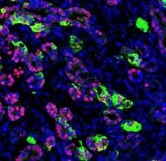
Insulin-Producing Pancreatic Cells Created from Human Skin Cells

Original article written by Dana Smith for Gladstone Institutes on January 6, 2016. Click here to read the original article.

SAN FRANCISCO, CA—Scientists at the Gladstone Institutes and the University of California, San Francisco (UCSF) have successfully converted human skin cells into fully-functional pancreatic cells. The new cells produced insulin in response to changes in glucose levels, and, when transplanted into mice, the cells protected the animals from developing diabetes in a mouse model of the disease.

The new study, published in Nature Communications, also presents significant advancements in cellular reprogramming technology, which will allow scientists to efficiently scale up pancreatic cell production and manufacture trillions of the target cells in a step-wise, controlled manner. This accomplishment opens the door for disease modeling and drug screening and brings personalized cell therapy a step closer for patients with diabetes.

Pancreatic cells

Functioning human pancreatic cells after they’ve been transplanted into a mouse. [Image: Saiyong Zhu]

“Our results demonstrate for the first time that human adult skin cells can be used to efficiently and rapidly generate functional pancreatic cells that behave similar to human beta cells,” says Matthias Hebrok, PhD, director of the Diabetes Center at UCSF and a co-senior author on the study. “This finding opens up the opportunity for the analysis of patient-specific pancreatic beta cell properties and the optimization of cell therapy approaches.”

In the study, the scientists first used pharmaceutical and genetic molecules to reprogram skin cells into endoderm progenitor cells—early developmental cells that have already been designated to mature into one of a number of different types of organs. With this method, the cells don’t have to be taken all the way back to a pluripotent stem cell state, meaning the scientists can turn them into pancreatic cells faster. The researchers have used a similar procedure previously to create heart, brain, and liver cells.

After another four molecules were added, the endoderm cells divided rapidly, allowing more than a trillion-fold expansion. Critically, the cells did not display any evidence of tumor formation, and they maintained their identity as early organ-specific cells.

The scientists then progressed these endoderm cells two more steps, first into pancreatic precursor cells, and then into fully-functional pancreatic beta cells. Most importantly, these cells protected mice from developing diabetes in a model of disease, having the critical ability to produce insulin in response to changes in glucose levels.

“This study represents the first successful creation of human insulin-producing pancreatic beta cells using a direct cellular reprogramming method,” says first author Saiyong Zhu, PhD, a postdoctoral researcher at the Gladstone Institute of Cardiovascular Disease. “The final step was the most unique—and the most difficult—as molecules had not previously been identified that could take reprogrammed cells the final step to functional pancreatic cells in a dish.”

Sheng Ding, PhD, a senior investigator in the Roddenberry Stem Cell Center at Gladstone and co-senior author on the study, adds, “This new cellular reprogramming and expansion paradigm is more sustainable and scalable than previous methods. Using this approach, cell production can be massively increased while maintaining quality control at multiple steps. This development ensures much greater regulation in the manufacturing process of new cells. Now we can generate virtually unlimited numbers of patient-matched insulin-producing pancreatic cells.”

Holger Russ, PhD, was a co-first author on the paper from UCSF. Other Gladstone investigators include Xiajing Wang, Mingliang Zhang, Tianhua Ma, Tao Xu, and Shibing Tang. Funding was provided by the Roddenberry Foundation, National Institutes of Health, National Heart, Lung, and Blood Institute, National Eye Institute, National Institute of Child Health and Human Development, National Institute of Mental Health, California Institute of Regenerative Medicine, Prostate Cancer Foundation, and the Leona M. & Harry B. Helmsley Charitable Trust.

About the Gladstone Institutes

To ensure our work does the greatest good, the Gladstone Institutes focuses on conditions with profound medical, economic, and social impact—unsolved diseases of the brain, the heart, and the immune system. Affiliated with the University of California, San Francisco, Gladstone is an independent, nonprofit life science research organization that uses visionary science and technology to overcome disease.

Contact Person

Dana Smith
Direct line: 415.734.2532
Mobile: 415.806.6245

Learn More +

OUR PROJECTS

See our approved research projects and campaigns.

Role of the integrated stress response in type 1 diabetes pathogenesis
In individuals with type 1 diabetes (T1D), the insulin-producing beta cells are spontaneously destroyed by their own immune system. The trigger that provokes the immune system to destroy the beta cells is unknown. However, accumulating evidence suggest that signals are perhaps first sent out by the stressed beta cells that eventually attracts the immune cells. Stressed cells adapt different stress mitigation systems as an adaptive response. However, when these adaptive responses go awry, it results in cell death. One of the stress response mechanisms, namely the integrated stress response (ISR) is activated under a variety of stressful stimuli to promote cell survival. However, when ISR is chronically activated, it can be damaging to the cells and can lead to cell death. The role of the ISR in the context of T1D is unknown. Therefore, in this DRC funded study, we propose to study the ISR in the beta cells to determine its role in propagating T1D.
Wearable Skin Fluorescence Imaging Patch for the Detection of Blood Glucose Level on an Engineered Skin Platform
zhang
A Potential Second Cure for T1D by Re-Educating the Patient’s Immune System
L Ferreira
Validating the Hypothesis to Cure T1D by Eliminating the Rejection of Cells From Another Person by Farming Beta Cells From a Patient’s Own Stem Cells
Han Zhu
Taming a Particularly Lethal Category of Cells May Reduce/Eliminate the Onset of T1D
JRDwyer 2022 Lab 1
Can the Inhibition of One Specific Body Gene Prevent Type 1 Diabetes?
Melanie
Is Cholesterol Exacerbating T1D by Reducing the Functionality and Regeneration Ability of Residual Beta Cells?
Regeneration Ability of Residual Beta Cells
A Call to Question… Is T1D Caused by Dysfunctionality of Two Pancreatic Cells (β and α)?
Xin Tong
Novel therapy initiative with potential path to preventing T1D by targeting TWO components of T1D development (autoimmune response and beta-cell survival)
flavia pecanha