Final Project Update
We are excited to share our final update on generating and preserving human macular patches. Based on our preliminary data as well as new data collected during the first half of the DRC project, we were able to consistently revive photoreceptor light responses from research donor eyes recovered 3-4 hours postmortem or from organ donations after cardiac death where the eyes were typically recovered 0.5-1h postmortem. We also demonstrated that postmortem hypoxia is the dominant mechanism underlying the irreversible loss of the rod- and cone-mediated light responses during the postmortem enucleation delay using genetically engineered mice with either rod- (Gnat2-/-) or cone-dominant (Gnat1-/-) light responses. We submitted these data as a manuscript to Nature, where it is currently under review.
However, by the end of the first 6 months of the project, we had not yet been able to revive the synchronized activity of the inner retina, something that will be critical if we were to expect success for generating macular grafts for good vision restoration. Moreover, we had not yet demonstrated ex vivo conditions to preserve light signaling in the retina samples for 24 hours or more, which will be critical for performing ex vivo manipulations required to promote axon regeneration, which will be critical for phase II of this study. We are happy to announce that we made significant progress on these two important issues. Firstly, we established a critical postmortem time period of about 20 minutes for the eye recovery based on experiments using a pig as a model. Moreover, we confirmed the translation of this result to humans by establishing a new way of recovering the donor eyes from brain dead organ donors 1 – 20 minutes postmortem. In this way, we were able to revive the synchronized activity of the inner as measured by the ex vivo ERG b waves from peripheral and macular samples from four out of five donors so far. We also found that recovery of the ERG b waves in the macula requires faster ex vivo perfusion, something we speculate to be due to higher metabolic demand in the cone-dominant central retina compared to that in the rod-dominant peripheral retina. To our knowledge, this was the first time anyone has revived the ERG b waves in the macula from postmortem eyes, and we think that the presence of b waves may become the main biomarker for assessing the viability of the macular grafts and their suitability for transplantation. Secondly, we tested various conditions for preserving the light signaling in mice, pigs, and humans. We found that room temperature works better than cold (4oC) or warm (35oC). Perhaps the most important new finding was that preserving the b wave for more than 24 hours requires keeping the neural retina and retinal pigment epithelium together. In mice, storage in the eyecup or in the whole eye was superior compared to storing isolated neural retina samples. Dark rearing rather than light exposure led to better preservation of the retinal function. This was somewhat surprising as light should reduce the metabolic rate in the retina. We continue to optimize the storage conditions by testing other strategies for reducing metabolic rate, such as CNG and Ca2+ channel blockers. We are currently scaling up the research from organ donations after brain death both in San Diego and potentially in Utah in the future to complete Phase I of the project. We are extremely grateful for the donors and support from DRC, which has been critical for this project.
6-Month PROJECT UPDATE
We are excited to share the interim progress report of our project. This project’s primary goal is to determine the criteria and protocols for generating and maintaining viable macula patches from human donor eyes. The ultimate goal is to generate patches of the macula that, in the future, could be used to restore vision in blind type 1 diabetic patients. A first step to achieve this goal is to be able to recover patches of the human macula that can generate and transmit light signals from photoreceptors, the sensory neurons in the retina, all the way to ganglion cells, the output neurons in the retina that project their axons to the visual centers of the brain for visual perception. To do this, we have assembled a team that currently involves research scientists, clinicians, eye banks, and organ donor society. With this team, we have determined how time from death to eye recovery, cause of death, postmortem light exposure, hypoxia, and acidosis contribute to our ability to restore light-evoked responses in the retina and specifically in the human macula. We have established conditions for the ex vivo eyes. Normal retinal light signaling can be maintained for at least 20 hours, and ongoing experiments are expected to extend this time window significantly. Our results demonstrate that we can restore light-evoked photoreceptor responses under certain conditions when eyes are recovered 0.5 – 4 hours postmortem. However, the transmission of light signals from photoreceptors to second-order neurons is often compromised even in the eyes recovered just 30 minutes after death. Using mice in our experiments have demonstrated postmortem hypoxia as the driving force for this apparent irreversible loss of light signal transmission in the retina. Ongoing investigations are testing if conditioning of mice to hypoxia before death or postconditioning of the eyes using drugs to induce hypoxia signaling pathways could promote the revival of the light signal transmission in the postmortem eyes. On the other hand, in pigs, we are conducting experiments to determine the maximal time window from death to enucleation, still allowing a robust restoration of light signal transmission in the retina. Based on these experiments, our organ donor experiments’ criteria and protocols will be modified to establish a method for producing fully functional human macular patches.
PROJECT DESCRIPTION
One of the major complications in type 1 diabetes (T1D) is vision impairment. Diabetic retinopathy is the leading cause of blindness among working-age adults, and accounts for a high degree of disability and long-term morbidity. Current approaches to treat diabetic retinopathy have limited success to restore vision when the retina is damaged. We are investigating the feasibility and technical challenges associated with the far-reaching concept of retinal cell replacement, a future goal that we believe has the potential to improve vision to blind T1D’s.
We envision a retinal cell replacement program consisting of several stages with multi-disciplinary and multi-institutional clinicians and scientists. For this project, we want to address 2 phases: Phase 1 consists of maintaining the viability of retinal tissue in postmortem eyes. Phase 2 consists of identifying conditions that allow cells to integrate into the retina.
Over the past three years, we have generated preliminary data that supports the feasibility of Phase 1. With a multidisciplinary team consisting of representatives from regional eye banks, organ donor programs, visual chemists, electrophysiologists and clinicians, we have succeeded in developing the techniques for maintaining or reviving the viability of macular and peripheral retinal tissue in postmortem human eyes. Initially, we found the neuronal viability in the human macula from donor eyes was compromised. We attributed the poor outcomes to multiple obstacles, including prolonged death-enucleation times. To solve these problems, we adopted a novel approach, using human organ donor eyes.
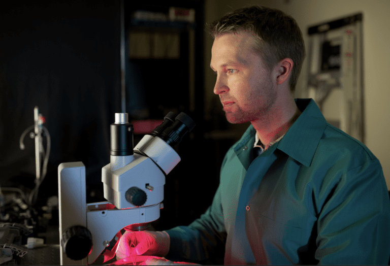
Dr. Hanneken became a registered organ donor research recipient and established a collaboration with LifeSharing, a major organ donor Society in San Diego and Imperial Valley. She harvested human organ donor eyes at the time that the transplant team was procuring heart and lung tissues, which overcame multiple obstacles. The responses with human organ donor eye tissues were significantly better, particularly in the outer retina where photoreceptor responses could be measured with good electrical amplitudes. However, there are still challenges and technical factors that reduce viability, particularly in the inner retina function.
In this project, we will use sophisticated approaches to examine the timing and the harvesting conditions of the human eye donor to optimize neuronal survival and/or revival of retinal neurons. These results are expected to establish criteria and protocols for potential retinal cell replacement capable of generating and transmitting light-evoked signals from photoreceptors all the way to the retinal ganglion cells, which are the output neurons of the retina.